This research, developed by the Wake Forest Baptist Medical Center in North Carolina, represents a breakthrough for regenerative medicine, it suggests that these structures could be implanted in patients in the future, overcoming “a number of technical obstacles” that hinder currently stand its leaders in a statement.
Experts printed “stable” and after implanting in rodents, matured into functional tissue, cartilage, bone and muscle structures while developed a system of blood vessels.
Although the new printed structures are not yet ready to be implanted in patients, remember, the first results of the study suggest that they have “the size, strength and proper functionality stop being used in humans. “
” This new printer tissues and organs is a major factor in our goal to manufacture replacement tissue for patients progress, “says Anthony Atala, director of the Institute for Regenerative Medicine at Wake Forest (WFIRM, its acronym in English).
According to the expert, the “bioprinter 3D” can be made “stable human scale of any shape and size tissue,” allowing “print living tissue and organ structures for surgical implantation “.
for this work, the WFIRM has been funded by the Institute for Regenerative Medicine of the US military, which aims to apply this technology in soldiers wounded in combat, given the shortage of donor tissue for implants.
the accuracy of this new 3D printer means that in the near future, could faithfully replicate more complex tissues and organs of the human body.
for now, remember the researchers current printers, whether injection laser or extrusion, can not reproduce structures having the size or strength necessary to be implanted in the body.
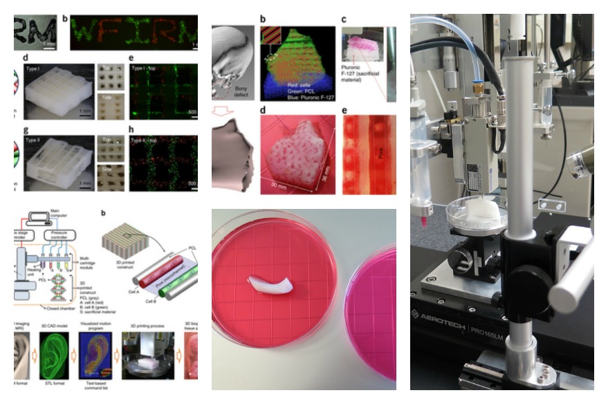
called Integrated Printing System Tissue and organ (ITOP), developed by the WFIRM during the last ten years, has overcome these limitations, held Atala.
the ITOP so much as biodegradable plastics to create the “form” of tissue and gels water-based holding cells.
in addition, the 3D machine manufactures a strong temporal external structure, which prevents cell damage occurring during the printing process.
Another of the challenges of tissue engineering is to make the implanted structures live long enough to be integrated into the body.
in this regard, experts optimized, on the one hand, “ink” of water based holding cells to improve their “health” and promote their growth, while printed a network of “micro” structures.
These channels point, allow nutrients and oxygen present in the human body are integrated in those structures, keep alive and develop a system of blood vessels.
previous research showed that cells survive only when the implanted tissue structures that have not been able to develop blood vessels have a size less than 200 microns (0.1778 mm).
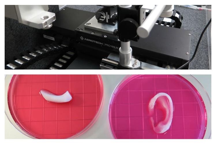
Atala and colleagues succeeded in making an ear of a size suitable for babies of 1.5 inches (38.1 millimeters) capable of survive and show signs of vascularization one two months after being implanted.
“Our results indicate that using a combined biotinta, with the development of micro, creates the right environment to keep cells alive and to encourage their growth and tissues” says the researcher.
Another feature of the ITOP is its ability to process data from scans and MRIs and “make tissue to measure” for each patient.
No comments:
Post a Comment